Physiotherapeut/in (m/w/d)
Gesucht wird ein/e motivierte/r
Physiotherapeut/in (m/w/d), der/die
unser tolles Team verstärkt und
sich auf eine abwechslungsreiche
und anspruchsvolle Tätigkeit in
einer modernen Praxis freut.
Deine Aufgaben:
Durchführung von individuellen
physiotherapeutischen Behandlungen
auf Grundlage einer ärztlichen
Verordnung
Erstellung von Therapieplänen und
Dokumentation der Behandlungen
Beratung und Anleitung von
Patienten zur Eigenübung und
Prävention
Eng...
Ich würde gerne wissen, ist es möglich, isoliert das Leistenband zu dehnen?
Sowie das ISG zu mobilisieren.
Was haltet ihr im Zusammenhang damit vom Vorlauftest als validen Test.
Danke für eure Antworten!
Gefällt mir
Wollen Sie diesen Beitrag wirklich melden?
Problem beschreiben
kinzi schrieb:
Hallo Schwarmwissen,
Ich würde gerne wissen, ist es möglich, isoliert das Leistenband zu dehnen?
Sowie das ISG zu mobilisieren.
Was haltet ihr im Zusammenhang damit vom Vorlauftest als validen Test.
Danke für eure Antworten!
Alexander M. Dydyk 1 , Stephen D. Forro 2 , Andrew Hanna 3
In: StatPearls [Internet]. Treasure Island (FL): StatPearls Publishing; 2024 Jan.
2023 Jul 4.
Affiliations
PMID: 32491804
Bookshelf ID: NBK557881
Free Books & Documents
Excerpt
Sacroiliac (SI) joint injury is a common cause of low back pain. Posterior pelvic joint pain a common name for SI joint dysfunction. The spine and pelvis are connected by the sacroiliac joint. The SI joint lies between the iliac's articular surface and the sacral auricular surface. When an injury occurs to the SI joint, patients often experience significant pain in their low back and buttock region. The SI joint experiences forces of shearing, torsion, rotation, and tension. Ambulation is heavily influenced by the SI joint, as this is the only orthopedic joint connecting our upper body to our lower body. The joint is a relatively stiff synovial joint filled with synovial fluid. The bones of the sacrum and ilium are coated in hyaline cartilage at their articular surfaces with dense fibrous tissue connecting the ilium and the sacrum. SI joints typically only have a few degrees of motion.
Diagnosing sacroiliac (SI) joint pathology can be challenging. One of the difficulties providers can run into in the evaluation of SI joint injury is in distinguishing between lower lumbar pain (lumbago) from SI joint pain. Specialized tests can be instrumental in making this distinction. It is vital to keep SI joint pain as part of the differential diagnosis of low back pain, with up to 30% of low back pain secondary to the SI joint. Pregnant women experience joint laxity due to hormonal changes, and this is when the SI joint is the most vulnerable to injury. Between the ages 40 and 50, the SI joint fuses decreasing the SI joints' laxity. Fusion and or pregnancy may lead to hypermobility or hypomobility, which may exacerbate SI joint pain. Osteoarthritis is a common cause of SI joint pain.
However, there are multiple etiologies and a variety of factors that can contribute to SI joint injury. The overlap in symptoms with various causes of low back pain, as well as the numerous origins of SI joint dysfunction, make it not only a tough diagnose to make but also challenging to treat. SI joint injury can be acute but becomes chronic after three months of persistent pain. Chronic SI joint pain occurs when the free nerve endings within the SI joint degenerate or become chronically activated. Pain can be constant or intermittent for SI joint injury. Given the prevalence of mechanical back pain, it is essential to rule out or exclude a lumbar origin of suspected SI joint pain before diagnosis. It is often hard to pinpoint in which cases SI joint dysfunction is the primary reason behind a patient's back pain. Some exceptions include trauma and pregnancy.
Extra joint mobility of the joint can result in pain in SI joint injury. However, hypomobility is a hallmark of ankylosing spondylitis, a common cause of inflammatory sacroiliac injury. SI joint dysfunction often occurs in unison with mechanical back pain. The sacroiliac joint may also be the site of pain referred from the lumbar vertebra rather than the origin of the patient's pain. For example, degenerative disc disease at the L5-S1 vertebrae may become interpreted at the SI joint, but the source is much higher in the lumbar spine. There are multiple patterns of referral of pain for patients with SI joint injury. Including, the posterior thigh, knee, or foot. The most common site of pain referral is the posterior thigh, seen in 50% of patients. Complicating SI joint injury management further is a lack of clearly defined guidelines in the diagnosis and management of SI joint pain. MRI is the test of choice in the evaluation of SI joint dysfunction. Furthermore, radiographic-guided anesthetic injection provides a reliable way of determining SI joint pain in many cases.
The sacroiliac joint is a commonly targeted area of treatment of chronic low back pain as well. Conservative treatment options for SI joint injury often include physical therapy, home exercises, over the counter pain medication such as NSAIDs or acetaminophen. When conservative management fails, corticosteroid injections and radiofrequency ablative therapies are viable treatment options. In severe, refractory cases, surgery can fuse the SI joint. Patient education is essential in SI joint injury, including posture, proper lifting technique, stretching, and regular exercise. Weight loss helps SI joint pain, as well.
Copyright © 2024, StatPearls Publishing LLC.
PubMed Disclaimer
Gefällt mir
Welche MT Tests würdest du empfehlen?
Und sollten 3 positiv sein, damit isg bestätigt?
Gefällt mir
Wollen Sie diesen Beitrag wirklich melden?
kinzi schrieb:
Vielen Dank für deine ausführliche Antwort!
Welche MT Tests würdest du empfehlen?
Und sollten 3 positiv sein, damit isg bestätigt?
Gefällt mir
Wollen Sie diesen Beitrag wirklich melden?
Geert Jeuring schrieb:
Hier gibt es die Laslett Teste und tatsächlich ist die Idee, das wenn 3 von 5 Teste positiv sind, es ein begründeten Verdacht auf eine Irritation des ISG-Gelenks gibt.Cluster of Laslett | Iliosakralgelenkschmerzen | SIJ Assessment
Wollen Sie diesen Beitrag wirklich melden?
Problem beschreiben
Geert Jeuring schrieb:
Hallo Kinzi, wenn es mit dem Englisch klappt, findest du hier Antworten, einiges habe ich versucht zu unterstreichen, aber das hat nicht geklappt: Kurzfassung: nur paar Grad Mobilität, Hypomobilität vor allem bei Bechterew, Hypermobilität vor allem bei Schwangeren, Reliabele Tests: MRT und radiologisch gesteuerte Injektionen. Auch Lasslett´s 5 Teste werden hier nicht erwähnt.
Alexander M. Dydyk 1 , Stephen D. Forro 2 , Andrew Hanna 3
In: StatPearls [Internet]. Treasure Island (FL): StatPearls Publishing; 2024 Jan.
2023 Jul 4.
Affiliations
PMID: 32491804
Bookshelf ID: NBK557881
Free Books & Documents
Excerpt
Sacroiliac (SI) joint injury is a common cause of low back pain. Posterior pelvic joint pain a common name for SI joint dysfunction. The spine and pelvis are connected by the sacroiliac joint. The SI joint lies between the iliac's articular surface and the sacral auricular surface. When an injury occurs to the SI joint, patients often experience significant pain in their low back and buttock region. The SI joint experiences forces of shearing, torsion, rotation, and tension. Ambulation is heavily influenced by the SI joint, as this is the only orthopedic joint connecting our upper body to our lower body. The joint is a relatively stiff synovial joint filled with synovial fluid. The bones of the sacrum and ilium are coated in hyaline cartilage at their articular surfaces with dense fibrous tissue connecting the ilium and the sacrum. SI joints typically only have a few degrees of motion.
Diagnosing sacroiliac (SI) joint pathology can be challenging. One of the difficulties providers can run into in the evaluation of SI joint injury is in distinguishing between lower lumbar pain (lumbago) from SI joint pain. Specialized tests can be instrumental in making this distinction. It is vital to keep SI joint pain as part of the differential diagnosis of low back pain, with up to 30% of low back pain secondary to the SI joint. Pregnant women experience joint laxity due to hormonal changes, and this is when the SI joint is the most vulnerable to injury. Between the ages 40 and 50, the SI joint fuses decreasing the SI joints' laxity. Fusion and or pregnancy may lead to hypermobility or hypomobility, which may exacerbate SI joint pain. Osteoarthritis is a common cause of SI joint pain.
However, there are multiple etiologies and a variety of factors that can contribute to SI joint injury. The overlap in symptoms with various causes of low back pain, as well as the numerous origins of SI joint dysfunction, make it not only a tough diagnose to make but also challenging to treat. SI joint injury can be acute but becomes chronic after three months of persistent pain. Chronic SI joint pain occurs when the free nerve endings within the SI joint degenerate or become chronically activated. Pain can be constant or intermittent for SI joint injury. Given the prevalence of mechanical back pain, it is essential to rule out or exclude a lumbar origin of suspected SI joint pain before diagnosis. It is often hard to pinpoint in which cases SI joint dysfunction is the primary reason behind a patient's back pain. Some exceptions include trauma and pregnancy.
Extra joint mobility of the joint can result in pain in SI joint injury. However, hypomobility is a hallmark of ankylosing spondylitis, a common cause of inflammatory sacroiliac injury. SI joint dysfunction often occurs in unison with mechanical back pain. The sacroiliac joint may also be the site of pain referred from the lumbar vertebra rather than the origin of the patient's pain. For example, degenerative disc disease at the L5-S1 vertebrae may become interpreted at the SI joint, but the source is much higher in the lumbar spine. There are multiple patterns of referral of pain for patients with SI joint injury. Including, the posterior thigh, knee, or foot. The most common site of pain referral is the posterior thigh, seen in 50% of patients. Complicating SI joint injury management further is a lack of clearly defined guidelines in the diagnosis and management of SI joint pain. MRI is the test of choice in the evaluation of SI joint dysfunction. Furthermore, radiographic-guided anesthetic injection provides a reliable way of determining SI joint pain in many cases.
The sacroiliac joint is a commonly targeted area of treatment of chronic low back pain as well. Conservative treatment options for SI joint injury often include physical therapy, home exercises, over the counter pain medication such as NSAIDs or acetaminophen. When conservative management fails, corticosteroid injections and radiofrequency ablative therapies are viable treatment options. In severe, refractory cases, surgery can fuse the SI joint. Patient education is essential in SI joint injury, including posture, proper lifting technique, stretching, and regular exercise. Weight loss helps SI joint pain, as well.
Copyright © 2024, StatPearls Publishing LLC.
PubMed Disclaimer
Gefällt mir
Gefällt mir
Wollen Sie diesen Beitrag wirklich melden?
kinzi schrieb:
Da ging es mir auch nur um das grundsätzliche...ob man es überhaupt dehnen kann....
WEnn man einige Bäuche sieht, würde ich sagen ja......
Gefällt mir
Wollen Sie diesen Beitrag wirklich melden?
Geert Jeuring schrieb:
@kinzi
WEnn man einige Bäuche sieht, würde ich sagen ja......
Schmerz entsteht nicht immer dort, wo es wehtut! mfg hgb blush
Gefällt mir
Wollen Sie diesen Beitrag wirklich melden?
hgb schrieb:
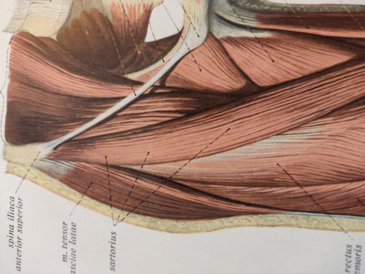
@kinzi ...das Lig. ist ein straffes Band, das aus den Aponeurosen der Bauchmuskeln und Strängen der fascia late entspringt, siehe hier: Ligamentum inguinale - DocCheck Flexikon  . Damit entfällt wohl die Dehnung des Bandes zu gunsten der Mobilisation der gen. Aponeurosen. Der Beckenring ist ein allseits dynamisches, in sich mobiles Gebilde, n. m. Erfahrung spielt das SIG keine so große Rolle, sondern das Ungleichgewicht der muskulären Steuerung. So helfen Abnehmen, Bauchtanz, Hula-Hoop und Beckenrollen wie Bebo-Gymnastik oder heute in hiesiger Zeitung "regelmäßige" Spaziergänge.
Schmerz entsteht nicht immer dort, wo es wehtut! mfg hgb blush
hat es Auswirkungen auf die Kniestabilität?
Gefällt mir
Wollen Sie diesen Beitrag wirklich melden?
kinzi schrieb:
@hgb
hat es Auswirkungen auf die Kniestabilität?
Gefällt mir
Wollen Sie diesen Beitrag wirklich melden?
kinzi schrieb:
@Geert Jeuring joy
Gefällt mir
Wollen Sie diesen Beitrag wirklich melden?
kinzi schrieb:
@Geert Jeuring ich meinte gezielt therapeutisch...
Gefällt mir
Wollen Sie diesen Beitrag wirklich melden?
hgb schrieb:
@kinzi Alle Muskeln,die am Becken ansetzen, beeinflussen sich gegenseitig. Rectus fem. Irritationen können die Kniestabilität beeinflussen, chrakteristisch treppab! Ein nicht ausreichend zu verlängernder Rectus wird gern wegen der zwei Gelenke übersehen, Test in BL und Ferse zum Gesäß im Seitvergleich.
Also, ich denke die Kräfte die dafür notwendig wären, würden als Körperverletzung gelten. Und um es mal sokratisch an zu gehen: was wäre der Therapeutische Sinn?
Gefällt mir
Wollen Sie diesen Beitrag wirklich melden?
Geert Jeuring schrieb:
@kinzi
Also, ich denke die Kräfte die dafür notwendig wären, würden als Körperverletzung gelten. Und um es mal sokratisch an zu gehen: was wäre der Therapeutische Sinn?
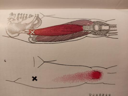
im Triggerhandbuch gibt es folgende Bilder bzw.Text:
Danach liegt der ob. TrP in der gen. Region "Leistenband". Travell hat immer nur die Schmerzpunkte gelistet und nur ab und zu auf vegetative Symptome wie Schwindel oder Arrhythmie des Herzens hingewiesen. Ein TrP wie dieser kann also durchaus ohne
störenden Schmerz die Stabilisierung des rectus fem beeinträchtigen.
D.h. der rectus sollte dort und auf Verlängerungsfähigkeit untersucht werden
Zu diesem TrP noch der zug. Text. mfg hgbblush
Gefällt mir
Wollen Sie diesen Beitrag wirklich melden?
hgb schrieb:
@kinzi


im Triggerhandbuch gibt es folgende Bilder bzw.Text:
Danach liegt der ob. TrP in der gen. Region "Leistenband". Travell hat immer nur die Schmerzpunkte gelistet und nur ab und zu auf vegetative Symptome wie Schwindel oder Arrhythmie des Herzens hingewiesen. Ein TrP wie dieser kann also durchaus ohne
störenden Schmerz die Stabilisierung des rectus fem beeinträchtigen.
D.h. der rectus sollte dort und auf Verlängerungsfähigkeit untersucht werden
Zu diesem TrP noch der zug. Text. mfg hgbblush
Gefällt mir
Wollen Sie diesen Beitrag wirklich melden?
kinzi schrieb:
@Geert Jeuring Mein Chef meint zur Beeinflussung der Kniestabilität
Gefällt mir
Wollen Sie diesen Beitrag wirklich melden?
kinzi schrieb:
@hgb naja, ich finde es ehrlich gesagt doch etwas weit weg vom Leistenband....
Gefällt mir
Wollen Sie diesen Beitrag wirklich melden?
kinzi schrieb:
Und hat es zur Demo an mir auch "gedehnt"....
.. das ist Dein gutes Recht, ich halte mich an die Anatomie!
Gefällt mir
Wollen Sie diesen Beitrag wirklich melden?
hgb schrieb:
@kinzi


.. das ist Dein gutes Recht, ich halte mich an die Anatomie!
Gefällt mir
Wollen Sie diesen Beitrag wirklich melden?
hgb schrieb:


Gefällt mir
Wollen Sie diesen Beitrag wirklich melden?
Geert Jeuring schrieb:
@kinzi
kinzi schrieb am 21.06.2024 22:41 Uhr:@Geert Jeuring Mein Chef meint zur Beeinflussung der Kniestabilität Wie soll denn das funktionieren? Kniestabilität in der Frontalebene findet vor allem durch Außenrotatoren und Adduktoren der Hüfte, so wie Tib. post und ant. statt, in der saggitale Ebenen durch Quadriceps und Ischiocruralgruppe. Das sollte dein Chef dir noch mal näher erklären.
Gefällt mir
Wollen Sie diesen Beitrag wirklich melden?
hgb schrieb:
jaja, der proximale Quadriceps, setzt er nicht am Hirn an??joy
Das denke ich auch. Ich bin da jetzt seit 1 Monat in der Praxis. Er möchte der beste Physio der Stadt werden/sein und ich dachte, ich kann noch viel von ihm lernen und ich weiß nicht, was ich von all dem halten soll...Elektrobehandlung en masse, Kinesiologische Abfrage von Organfunktionen, ISG Vorlauftest als Standartuntersuchung bei Lumbargo und dann solche Aussagen wie oben.ui ui ui...confused
Gefällt mir
Wollen Sie diesen Beitrag wirklich melden?
kinzi schrieb:
@Geert Jeuring
Das denke ich auch. Ich bin da jetzt seit 1 Monat in der Praxis. Er möchte der beste Physio der Stadt werden/sein und ich dachte, ich kann noch viel von ihm lernen und ich weiß nicht, was ich von all dem halten soll...Elektrobehandlung en masse, Kinesiologische Abfrage von Organfunktionen, ISG Vorlauftest als Standartuntersuchung bei Lumbargo und dann solche Aussagen wie oben.ui ui ui...confused
Irgendwie hast du kein Glück mit deinen Chefs. Warst du nicht erst vorher in einer Praxis mit schlechter Bezahlung?
Gefällt mir
Wollen Sie diesen Beitrag wirklich melden?
sabine963 schrieb:
@kinzi
Irgendwie hast du kein Glück mit deinen Chefs. Warst du nicht erst vorher in einer Praxis mit schlechter Bezahlung?
Gefällt mir
Wollen Sie diesen Beitrag wirklich melden?
kinzi schrieb:
@sabine963 Das stimmt....nun stimmt die Bezahlung, aber dann so was....
Gefällt mir
Wollen Sie diesen Beitrag wirklich melden?
pt ani schrieb:
@kinzi Dann mach Du es so gut, wie Du kannst. Das spricht sich schnell herum.
Wollen Sie diesen Beitrag wirklich melden?
Problem beschreiben
Geert Jeuring schrieb:
Zu dem Leistenband noch: das hat wohl eher weniger mit Stabilität oder Mobilität des Beckens zu tun sondern wird gebildet aus oder bietet Stutz an verschiedene Strukturen u.a. die Bauchmuskulatur.
Mein Profilbild bearbeiten